In brief — the direct answer
Placenta praevia covers the cervical opening, while placenta accreta spectrum involves abnormal placental attachment or invasion; the conditions can occur together but are not the same diagnosis. Both may cause serious bleeding. Assessment guides an individual delivery plan, which may require a specialist multidisciplinary team, blood-bank support and counselling about surgical risks. Planning reduces selected risks but cannot guarantee safety.
What is Placenta Previa & Accreta Spectrum?
The conditions are related to how the placenta implants early in pregnancy:
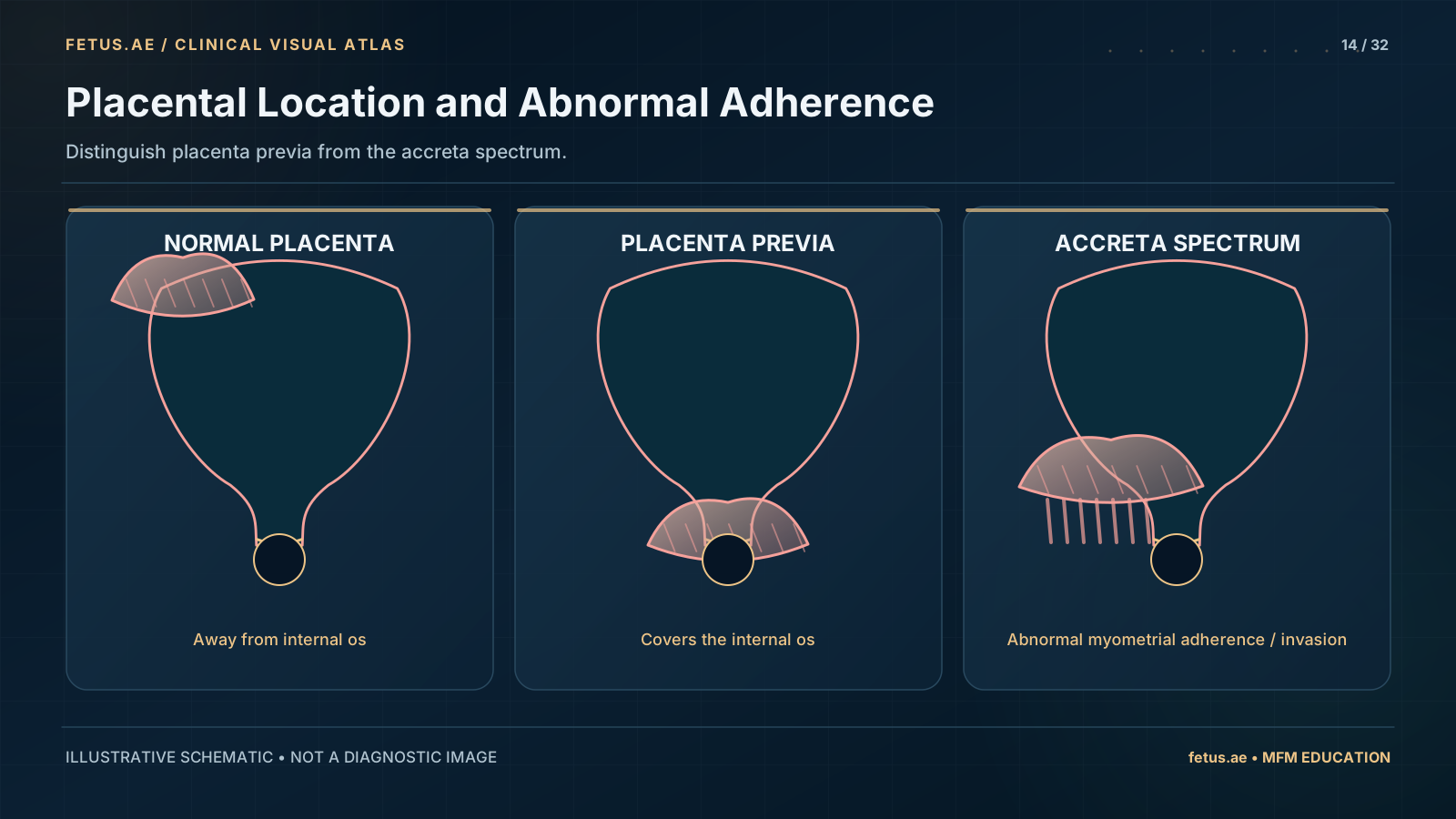
- Placenta Previa: The placenta is 'low-lying.' As the cervix begins to open or thin late in pregnancy, the placenta's blood vessels can tear, causing sudden, painless bleeding.
- Placenta Accreta Spectrum (PAS): This includes Accreta, Increta, and Percreta. Here, the placenta attaches so firmly that it cannot be removed naturally after the baby is born. In the most severe form (Percreta), the placenta may sogar grow through the uterus and attach to nearby organs like the bladder.
 Enlarge schematic
Distinguish placenta previa from the accreta spectrum.
Simplified educational schematic; not to scale or for diagnosis.
Who is at risk?
The most significant risk factor is a history of prior C-sections. The more C-sections a woman has had, the higher her risk of developing Accreta, especially if the current placenta is implanted over the old scar. Other factors include previous uterine surgeries (like myomectomy for fibroids) and smoking.
How we diagnose it
Diagnostics focus on high-fidelity imaging:
- Expert Ultrasound: Using specialized Doppler to look for 'lacunae' (holes in the placenta) and the loss of the 'clear zone' between the placenta and the uterus.
- Fetal MRI: In complex cases, we use MRI to assess exactly how deeply the placenta has invaded the uterine wall or if it has reached the bladder.
- Early Screening: Placenta position is checked at the 20-week anomaly scan and followed closely if it is low-lying.
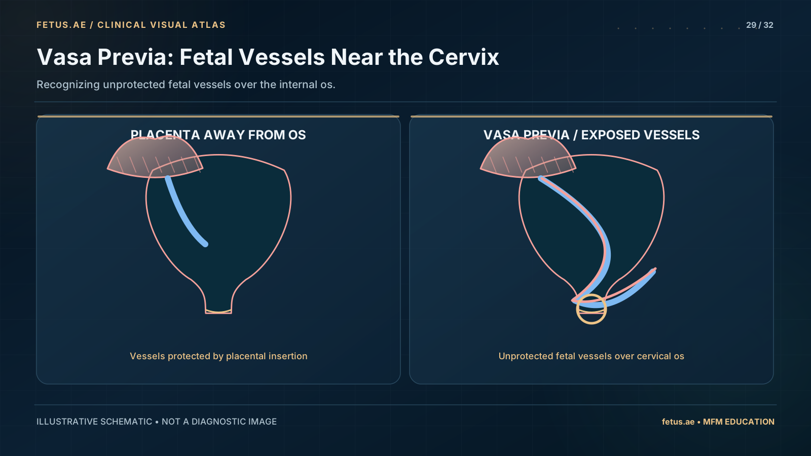
Related placental condition: vasa praevia
 Enlarge schematic
Recognizing unprotected fetal vessels over the internal os.
Simplified educational schematic; not to scale or for diagnosis.
What does management involve?
The management of PAS is one of the most complex tasks in obstetrics:
- Planned Delivery: We usually deliver by scheduled C-section between 34 and 36 weeks of pregnancy.
- Multidisciplinary Team: Our surgical team includes MFM specialists, urologists, vascular surgeons, and expert anesthesiologists.
- Blood Conservation: We use specialized equipment (cell-savers) to recycle a woman's own blood and have a large supply of blood products ready if needed.
- Cesarean Hysterectomy: In many cases of confirmed PAS, the safest path is to deliver the baby and then remove the uterus with the placenta still attached to prevent life-threatening hemorrhage.
What are the outcomes?
With expert planning, the outcomes for mothers and babies are excellent:
- Safety: By transforming an 'emergency' hemorrhage into a 'planned' surgical procedure, we significantly reduce the risks of severe blood loss.
- The Baby: While the baby may be born a few weeks early, they typically do very well after a short stay in the NICU.
- Recovery: Recovery from a PAS surgery is more intensive than a standard C-section, requiring specialized post-operative care in a high-dependency unit.
When to see a subspecialist
If your ultrasound mentions 'placenta previa' or 'placenta accreta,' or if you have any painless bleeding in the second or third trimester, you must be referred to a tertiary center.
Our Unit, led by Dr. Gareth Waring and Dr. Leanne Bricker, provides the specialized imaging and surgical expertise required for PAS. We provide a 'one-stop' multidisciplinary approach, ensuring all the necessary surgeons and resources are in one place for your birth.